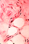
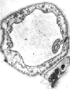
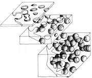
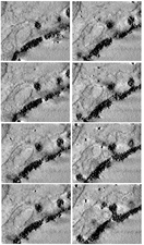
Three Dimensional Analysis of the Capillary Wall

with TEM Tomography

R. Wagner. S. Modla , F. Hossler & K. Czymmek

Dept. Biological Sciences &

Delaware Biotechnology Institute

University of Delaware


Transmission electron micrographs of thin sections through capillary endothelial cells lack three dimensionality and the continuity or discreetness of membraneous vesicular compartments is indeterminate. Structure in the third dimension can be revealed by TEM Tomography of semi-thick sections which contain a significant fraction of vesicular structures. Electron projections of thick sections are recorded at 1 degree increments through 180 degrees of tilt. A second tilt series is acquired at right angles to the first The resolution of "Dual Axis Tomograms" is significantly improved. A back projection algorithm can be used to convert the two dimensional images into a three dimensional representation of the original structure. Tracer-filled compartments can be modeled and rotated in any direction for enhanced appreciation of three-dimensional details. Free vesicles, larger membranous compartments connected to both luminal and abluminal surface and transendothelial channels of fused vesicles are revealed by TEM tomography.


singcap thinthick series models
Blood Capillaries Capillary Endothelium Thin vs Thick Sections TEM Tomography Computer Modeling